Table of Contents
Respiratory System of Branchiostoma:
In Branchiostoma respiration occur through the general surface area of metapleural folds, atrial wall, pharyngeal wall. The blood vessels present on the body wall and the oxygen present in the water current come close contact and gaseous exchange take place. The gill slits and gill bars have no special arrangements of blood vessels or capillaries so the gill does not take place a significant role in respiration.
The blood does not present any respiratory pigment, the respiratory gases diffused in the blood directly to different parts of the body.
Blood Vascular System of Branchiostoma:
Blood Vascular System is closed type in Branchiostoma, the blood is colourless due to absence of respiratory pigment in their blood. In Branchiostoma the blood does not have any blood cells or blood corpuscles, besides blood, they have lymph like body fluids in some portion of their body, as in the metapleural folds.
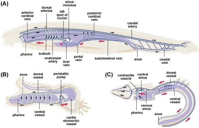
The heart is absent in the blood vascular system of Branchiostoma, the muscular blood vessels contract and provides the pumping force to the blood. Their blood vessels are not differentiated into arteries and veins, there are no structural differences between different blood vessels.
Sinus Venosus:
It is a sac-like structure present near the midgut diverticulum and pharynx on the ventral side of the body. All the blood from different parts of the body is collected by the sinus venosus through different veins. Sinus venosus collect and send the blood to the ventral aorta.
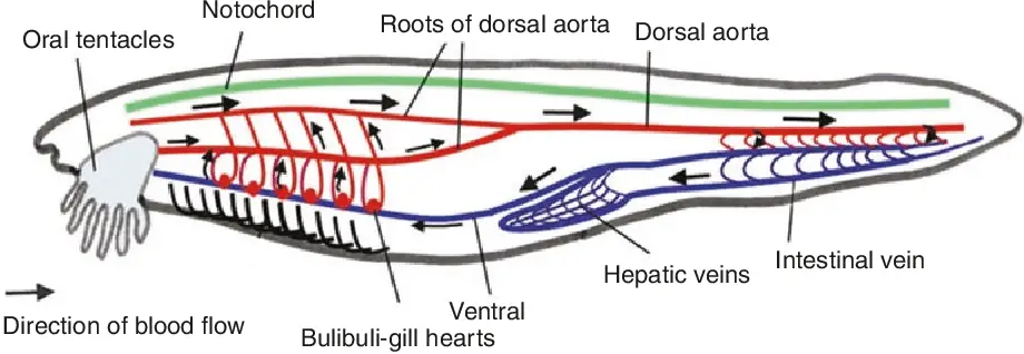
Ventral Aorta:
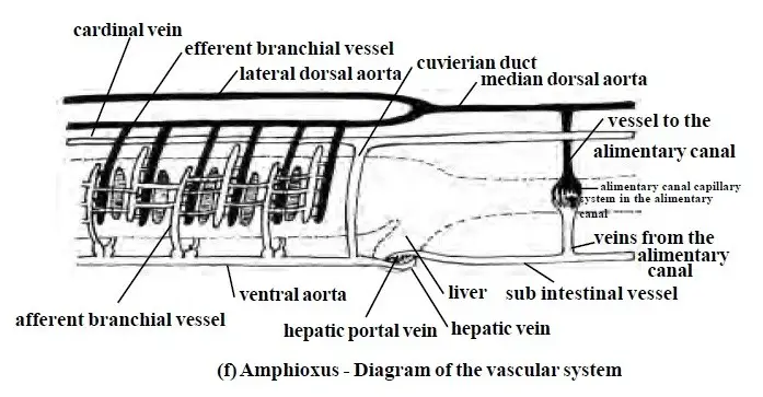
The ventral aorta runs in a forwarding direction along the mid-ventral line of the pharynx region through the ventral wall of the pharynx under the endostyle. The ventral aorta runs through the subendostylar region, for this reason, the ventral aorta is also known as subendostylar aorta. As there is no heart, the muscular contraction of the ventral aorta force the blood in a forwarding direction.
In the way of the ventral vessel, several lateral afferent branchial arteries arise in pairs and they supply blood to the primary gill bars. At the base of each primary gill bar, the branchial arteries produce a bulb-like structure known as bulbillus or bulbulli. All the afferent arteries present in primary gill bars are connected to secondary gill bars through transverse vessels of synapticulae.
Each afferent branchial arteries break down into a fine network of blood capillaries and form a nephric glomerulus sinus or glomus. The blood capillaries of the nephric glomerulus sinus finally join together and form efferent branchial arteries, the efferent arteries of the left side join into the lateral dorsal aorta on the left side and the efferent arteries of the right side open into the right lateral dorsal aorta.
Dorsal Aorta:
Two aortae are present on the left and right lateral sides of the pharynx known as the left lateral dorsal aorta and right lateral dorsal aorta. On the anterior region two lateral dorsal aortae join together and form an internal carotid artery that supplies blood to the oral hood region.
On the posterior region of the pharynx two lateral dorsal aortae join together and form a single median dorsal aorta. The dorsal aorta runs in between the notochord and intestine and it finally opens into the caudal artery in the tail region. The median dorsal aorta and two lateral dorsal aortae give some branches which supply blood to the myocoel region. Myocoel is the space between the myotomes are body walls.
Intestinal arteries arise from the median dorsal aorta and form a plexus of blood vessels in the intestinal wall. The blood flows in the median dorsal aorta in backwards directions while in the ventral aorta blood flow in the forwarding direction.
Sun-intestinal Veins:
On the ventral side below the intestinal, a plexus of blood vessels is presently known as the sun-intestinal vein. The sub-intestinal vein collects blood from the tail region through the mid-ventral caudal vein and collects blood from the intestine through lateral intestinal veins.
Hepatic Portal System:
The sun-intestinal vein on the anterior region forms the hepatic portal system in the midgut diverticulum region. The hepatic portal system breaks down into blood capillaries and forms a network of capillaries in the midgut diverticulum region. The hepatic portal system finally meets with the sinus venosus on the ventral side and the presence of the hepatic portal system is a superior feature of phylum Chordata.
Cardinal Veins:
Anterior and posterior cardinal veins collect blood from the two ventrolateral body walls and finally meet behind of pharynx known as common cardinal veins or ductus Cuvieri. The two ductus Cuvieri run in a downward direction through the Atrium and join to the sinus venosus on the ventral side.
Lymphatic System:
In Branchiostoma metapleural folds and the fins have open space in which colourless lymph like fluid pressure and all those fluid-filled space are collectively known as the lymphatic systems. The lymphatic system is responsible for a major gaseous exchange occurring in the body.
Reference
Detailed Information on
Characteristics Features of Subphylum Urochordata
Classification of Subphylum Urochordata
Examples of Subphylum Urochordata: Clavellina, Salpa, and Doliolum
Examples of Subphylum Urochordata
Branchiostoma Habitat and Geographical Distribution
External Morphology of Branchiostoma
Body Wall and Endoskeleton of Branchiostoma
Coelom and Movement in Branchiostoma

Hi Everyone!!! Welcome to Imaluop. Imaluop always try to learn some new and he want to share to other people. Here we will try to learn various topics on Science, specially on Biological Sciences.