The digestive system of Branchiostoma is a complete and long tube with digestive glands associated with the alimentary canal.
Table of Contents
Alimentary Canal:
As we know that the digestive system in Branchiostoma is complete and it starts from the mouth and ends in the anus. The diameter of the lumen of the digestive tract is not equal in all portions of the digestive tract.
Mouth:
The mouth is present ventrally below the anterior-most rostrum part, the mouth is a large oval aperture that is surrounded by a membranous structure known as an oral hood.
Oral Hood:
The anterior and lateral fold of the trunk forms a projection around the mouth opening which is known as oral hood.
Buccal Cirri:
Along the ventrolateral margin of the oral hood around 10 tentacles like slender structures present are known as buccal cirri. The buccal cirri have sensory papillae which are responsible for the perception of sensory stimulation. The number of buccal cirri increases with the age of Branchiostoma. The buccal cirri are stiff due to the presence of rods like endoskeletal structure (made of gelatinous connective tissue).
When food cum respiratory water current enters into the mouth, the buccal cirri form a sieve-like structure which prevents entry of unnecessary large particles in the buccal cavity.
Buccal Cavity or Vestibule:
The mouth opening leads into a funnel-shaped space surrounded by an oral hood known as a buccal cavity or vestibule.
Wheel Organ:
The epithelium of the oral hood has around 8 fingers like projection and all the finger-like projections make a wheel-like structure known as a wheel organ. Each finger-like projection is made of the alternate ciliated ridge and ciliated grove. The wheel organ is responsible for creating a whirling movement in the water and creating a water current.
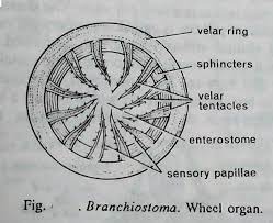
The ciliated epithelium of the wheel organ creates a whorling water current. The mid-dorsal large grove present on the roof of the buccal cavity is known as Hatschek’s groove. The mid-dorsal roof of the buccal cavity end into a small depression known as Hatschek’s pit and the pit is the responsible perception of sensory stimulation. The Hatschek’s groove and Hatschek’s pit are glandular, ciliated, and secret mucous.
Velum or Enterostome:
On the posterior side the vestibule or buccal cavity end to a vertical circular perforated membranous wall known as the vellum. The velum is perforated at the center, the circular central perforation on the velum is known as Enterostome. The Enterostome leads into the pharynx, some workers consider the Enterostome as mouth but it is not an actual mouth opening.
The inner border of the Enterostome is lined by a series of tentacle-like slender structures known as velar tentacles responsible for filtering the water current entering into the pharynx through the Enterostome. The opening of the Enterostome is controlled by a circular muscle sphincter present in the velar membrane.
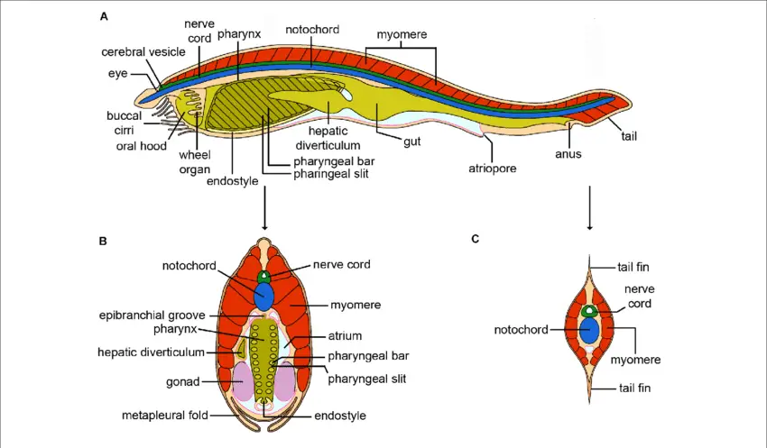
Pharynx:
The Enterostome leads into a large laterally compressed cavity in the alimentary canal which is actually the pharynx part. The pharynx is surrounded by an atrial cavity on the lateral and ventral sides but on the dorsal side, it does not surround by the atrial cavity.
Gill Slits:
On the lateral wall of the pharynx around 150 pairs of vertical gill slits are present through which the pharynx communicates with the atrial cavity. The number of gill slits increases with the age of the animals, the pharyngeal wall between two adjacent gill slits is known as the gill bar.
The gill bars are covered by ciliated epithelium and supported by an endoskeletal structure known as gill rods. The gill bars are of two types, primary gill bars and secondary gill bars, primary and secondary gill bars present alternately.
The gill rods present in the primary gill bar and secondary gill bar have a different structure, the skeletal rod present in the primary gill bar are bifurcated but in secondary gill bars the skeletal rod is not bifurcated, they are simple.
Endostyle:
It is a shallow ciliated grove along the mid-ventral line of the pharynx. On the ventral side, the endostyle is supported by two gelatinous plates.
Epipharyngeal Groove:
A ciliated groove present along the mid-dorsal line of the pharynx is known as the epipharyngeal groove. The epipharyngeal groove lead into the opening of the esophagus in the posterior region.
Oesophagus:
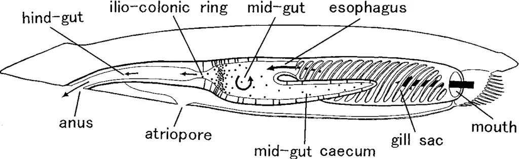
The posterior region of the pharynx leads into a tubular part of the alimentary canal known as the esophagus, the esophagus has ciliated epithelium.
Intestine:
The esophagus lead into the tubular intestinal part, the intestine or gut remains in a suspended state in the atrial cavity attached to the dorsal mesentery. The intestine has three different parts, the anterior wider part is known as the midgut and the terminal narrow part is known as the hindgut, the blind sac tubular part arises from the junction of the esophagus, and the midgut is known as the diverticulum. The diverticulum is present on the right lateral side of the alimentary canal lined by ciliated epithelium.
Anus:
The narrow hindgut opens outside through the anus, the circular small aperture of the anus is controlled by a muscular sphincter. The anus opens outside at the base of the caudal fin, slightly left to the mid-ventral line.
Reference
Detailed Information on
Characteristics Features of Subphylum Urochordata
Classification of Subphylum Urochordata
Examples of Subphylum Urochordata: Clavellina, Salpa, and Doliolum
Examples of Subphylum Urochordata
Branchiostoma Habitat and Geographical Distribution
External Morphology of Branchiostoma

Hi Everyone!!! Welcome to Imaluop. Imaluop always try to learn some new and he want to share to other people. Here we will try to learn various topics on Science, specially on Biological Sciences.