The body wall in Branchiostoma have three layers, from outside to inside the three layers are the outermost skin layer, middle muscular layer and inner parietal peritoneum.
Table of Contents
Skin:
It is the outermost thin delicate layer, this layer is made of a single layer of columnar epithelium cells. The columnar epithelium cells present on the basement membrane are attached to the membrane and on the outer surface a layer of cuticle formed as a result of secretion of epithelial tissue, the cuticle later interrupted in some places.
In young Branchiostoma the columnar epithelium cells have cilia but with the age, the epithelial cilia are lost and in adults, Branchiostoma cilia are absent on the columnar epithelium cells. The columnar epithelium tissue does not have any glandular or pigmented cells, this layer has sensory cells.
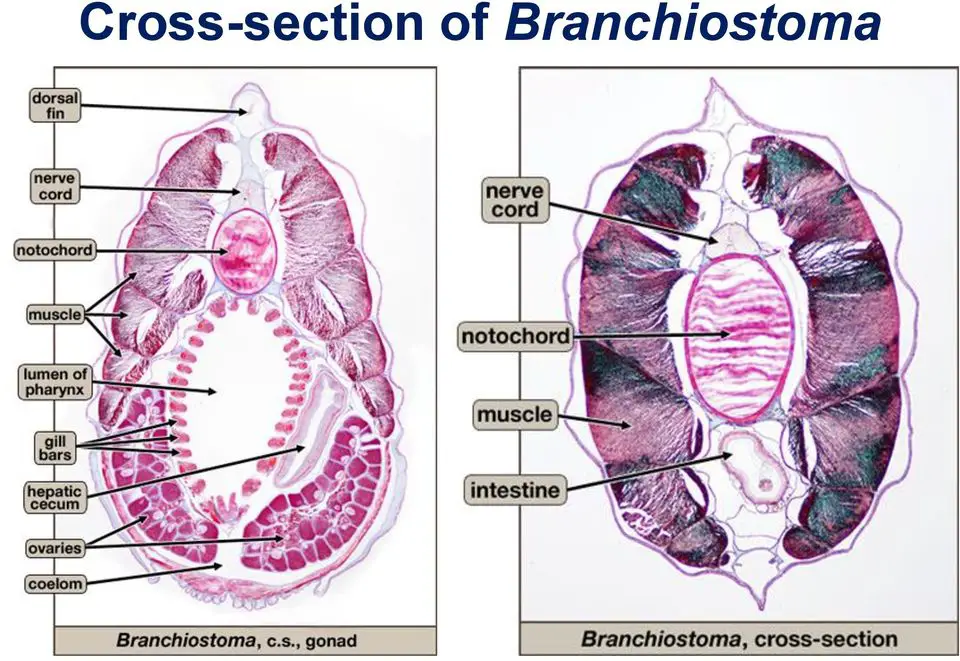
Below the epidermis, a dense layer is presently known as cutis, this layer is formed of fibrous connective tissue. Below the cutis layer, another gelatinous spongy layer is present which is known as subcutis, in this layer nerve fibres and blood vessels are present. Some cells present in the skin act similar functions as that of fibroblasts in higher Chordate, those cells are responsible for the formation of cutis and subcutis layer.
Muscle Layer:
In Branchiostoma special arrangements of muscle fibres are observed, along the whole length of their body on the dorsal side and later side muscle fibres arranged in several segments. On two lateral sides, they have V-shaped muscle fibres segments one after another, the V-shaped muscle segments are known as myotomes or myomeres. The apex of V-shaped myotomes is present in the anterior direction and the number of myotomes is around 60, but it very from species to species.
Each myotome is packed inside connective tissue covering which separates each myotome from one another, the connective tissue separator is known as myosepta or myocommata. In each myotome, striated muscle fibres are arranged in such a manner that the contraction of myotomes creates an undulation in the body. The lateral undulation movement in Branchiostoma helps them to swim in the water actively. On the ventral side, some transverse muscle fibres are arranged from two lateral metapleural folds, contraction of these muscle fibres force the water inside the atrium to exit through the atriopore.
Parietal Peritoneum:
It lines the inner boundary of the body wall, the parietal peritoneum separates the outer part of the body from the body wall. The parietal peritoneum is not present throughout the body because it is interrupted in some places.
Endoskeleton of Branchiostoma:
In Branchiostoma there is no exoskeleton, some endoskeletal structure is present but they are not cartilaginous or bony.
Notochord:
On the dorsal side notochord or chorda dorsalis present as a primary axial endoskeleton in Branchiostoma. In Branchiostoma the notochord is present throughout the length of the body from the tip of the rostrum to the end of the tail below the nerve cord and above the gut. The notochord is tapering at both ends and its extension beyond the brain up to the rostrum is a special feature in Branchiostoma.
The notochord is made of large vacuolated cells filled with fluid but in the adult stage, the notochord is made of gelatinous discs arranged in linear arrangements. The notochord is covered by a tough connective tissue membrane known as the notochordal sheath, the elasticity of the notochord make the animals bend.
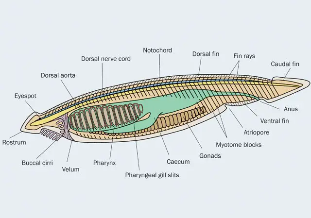
Fin-rays of Branchiostoma:
The dorsal and ventral fin is supported by a rectangular connective tissue box like fin-ray which supports the fin. The dorsal fin is supported by one row of gelatinous matrix-like fin-ray but on the ventral side two rays of ventral fin rays.
Oral Ring of Branchiostoma:
On the anterior side oral hood is supported by an endoskeletal structure made of connective tissue known as the oral ring. The part of the oral ring also supports the cirri of the oral hood.
Gill Rods in Branchiostoma:
The gill slits have gill bars, the gill bars are supported by a special type of endoskeletal structure known as gill rods. The gill rods are made of gelatinous connective tissue, in Branchiostoma two types of gill rods are present. One type of gill rod is bifurcated ventrally known as primary gill rods another type of gill rods are known as secondary gill rods.
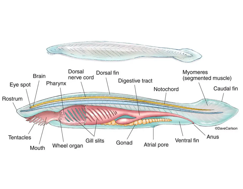
In Branchiostoma some special connective tissue is present below the epidermis which supports various visceral organs and the endostyle is supported by two endostylar plates.
Reference
Detailed Information on
Characteristics Features of Subphylum Urochordata
Classification of Subphylum Urochordata
Examples of Subphylum Urochordata: Clavellina, Salpa, and Doliolum
Examples of Subphylum Urochordata

Hi Everyone!!! Welcome to Imaluop. Imaluop always try to learn some new and he want to share to other people. Here we will try to learn various topics on Science, specially on Biological Sciences.