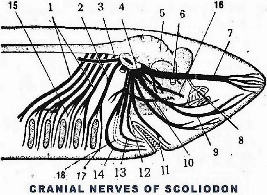
The peripheral Nervous System of Scoliodon includes the nerve that arises from the brain (cranial nerve) and the nerve that arises from the spinal cord (spinal nerve). In Scoliodon the brain gives 10 pairs of cranial nerves, the function of all cranial nerves is almost similar to that of the cranial nerve of the higher Vertebrate.
Table of Contents
Nerve O: Preolfactory Nerve:
All the cranial nerves are known by their specific name as well as the roman numerals, but the terminal cranial nerve in Scoliodon was discovered much after the discovery of other cranial nerves so the terminal cranial nerve has the name O. In the ventral side of the cerebellum through the neuropore thin terminal nerve arise which present in the olfactory region, this nerve is sensory in nature.
Nerve I: Olfactory Nerve:
The olfactory nerve takes part in understanding smell, the olfactory nerve arises from the olfactory lobe and passes through the olfactory sac.
Nerve II: Optic Nerve:
It is a sensory nerve important for the vision, arising from the optic thalamus on the ventral side of the diencephalon innervate to the retina of the eyes. The optic nerve after its origin crosses each other which forms a cross known as optic chiasma.
Nerve III: Oculomotor:
It is a motor nerve that controls the movement of eyeballs, iris, and lens, the oculomotor nerve arises from the ventral surface of the midbrain. After its origin, the oculomotor nerve is divided into four branches, the branches of the oculomotor nerve innervate the inferior rectus muscle, superior rectus muscle, anterior rectus muscle, and oblique muscle of the eye.
Nerve IV: Trochlear Nerve:
It is a motor nerve that also helps in the movement of the eyeball, the pathetic or trochlear muscle arising from the dorsolateral side of the midbrain. The trochlear nerve innervates the superior oblique muscle of the eyeball which controls the eye movements.
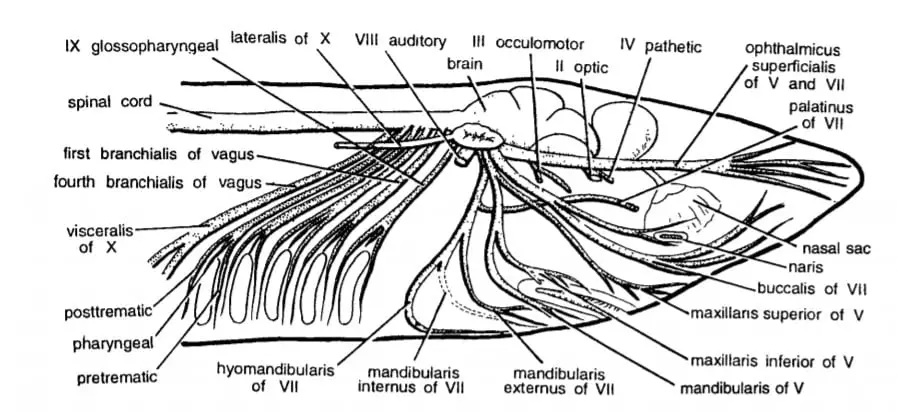
Nerve V: Trigeminal Nerve:
The trigeminal nerve is a mixed large nerve arising from the dorsolateral side of the anterior end of the medulla and it gives four branches. The trigeminal nerve inside the cranium shows a small ganglion known as Gasserian ganglion.
Opthalmicus Superficialis:
This branch proceeds in the forwarding direction and in the orbit region it intermingled with the VII nerve, reaches in the anterior region, and perceives the sensory stimulation from the skin of the snout.
Opthalmicus Profunds:
It is a sensory branch of the trigeminal nerve which innervates to the dorsal region of the snout, joining with superficial ophthalmic branch.
Maxillaries:
It carries the sensory stimulation from the ventral wall of the snout, it has two branches. One of them is maxillaris superior, it innervates ventral skin of the snout, in orbit region, it runs along with the buccal branch of the nerve VII, it is a flat ribbon-like nerve.
Maxillaris inferior is the branch of the maxillaris that innervates the posterior part of the upper jaw.
Mandibularis:
It is a mixed nerve that gives supply to the muscle of the lower jaw, tongue, and gill, it runs along the posterior wall of the orbit.
Nerve VI: Abducens Nerve:
It is a motor nerve innervated into the posterior rectus muscle of the eye, it originates in the midventral region of the medulla.
Nerve VII: Facial Nerve:
It is a fixed large-sized nerve arising from the lateral side of the medulla and innervating to the same region as that of the trigeminal nerve. The facial nerve has four branches – opthalmicus superficialis, buccalis, palatinus, hyomandibular.
Opthalmicus Superficialis:
It innervates to the upper side of the orbit and ampullae of Lorenzini along with the opthalmicus superficialis branch of the trigeminal nerve.
Buccalis:
Buccalis and maxillaris superior branch of trigeminal nerve together form a common infraorbital nerve which runs along the floor of the orbit, this branch supplies the ampullae of Lorenzini and the infraorbital canal.
Palatinus:
This branch of the facial nerve innervates the roof of the buccal cavity and pharyngeal cavity, it runs along the floor of the orbit.
Hyomandibular:
It has further three branches, hyomandibular branch runs along the posterior wall of the orbit.
- Hyoidean branch supply to the muscle of the hyoid arch or throat.
- Mandibularis internus branch give supply to the mucous membrane of the buccal cavity.
- Mandibularis externus is the nerve beach which innervate to the mandibular canal.
Nerve VIII: Auditory Nerve:
It is a thick and short sensory nerve that innervates to the membranous labyrinth of the auditory capsule and carries the sound wave to the brain.
Nerve IX: Glossopharyngeal Nerve:
It is a mixed nerve that arises from the ventrolateral region of the medulla and is divided into three branches – pretrematic branch runs along the anterior side of the first-gill pouch, posttrematic branch run along the posterior side of the first-gill pouch, pharyngeal branch innervates to the mucous membrane of the pharynx.
Nerve X: Vagus Nerve:
It is the largest cranial nerve in Scoliodon that arises from the posterior lateral side of the medulla, it has three branches.
- Lateralis: On lateral side it run along the lateral line canal and innervate the sensory structure in lateral line.
- Visceralis: This nerve branch supply to different visceral organs of the body cavity, like it innervate the heart, digestive system.
- Branchialis: Total four branchialis nerve supply to the four gill pouches, second to fivth gill pouches. Each branchialis again have three branches, pretrematic along the anterior side of gill pouch, posttrematic along the posterior side of the gill pouch, pharygeal supply to the mucous membrane of pharynx.
Spinal Nerve:
Along the entire length of the spinal cord, spinal nerves arise after certain regular intervals are known as spinal nerves. Each spinal nerve has a sensory dorsal root and motor ventral root, the sensory dorsal root is ganglionated.
Outside the neural arch, the dorsal root and the ventral root join together to form a mixed nerve, and each spinal nerve show three branches.
- The ramus dorsalis branch of each spinal nerve in Scoliodon innervate the muscle and skin of dorsal body wall.
- The ramus ventralis branch or each spinal nerve in Scoliodon innervate the muscle and skin of the ventral body wall.
- The ramus communicans branch of the spinal nerve connect with the autonomous nervous system.
In the pectoral region, a branchial Nerve plexus is present but in the pelvic region, no such nerve plexus is present in Scoliodon.
Detailed Study On
Central Nervous System of Scoliodon
Respiratory System of Scoliodon
Digestive Glands and Feeding Mechanism in Scoliodon
Coelom and Viscera in Scoliodon
Placoid Scales In Scoliodon (Exoskeleton)
Skin of Scoliodon (Integument)

Hi Everyone!!! Welcome to Imaluop. Imaluop always try to learn some new and he want to share to other people. Here we will try to learn various topics on Science, specially on Biological Sciences.