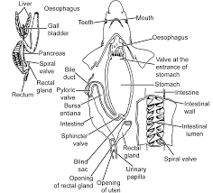
The Alimentary Canal of Scoliodon starts at their ventral slit-like transverse mouth opening which leads into the dorso-ventrally flattened buccal cavity. The buccal cavity opens into the large cavity of the pharynx, the pharynx opens into a short narrow esophageal on the posterior end. The esophagus opens into the U-shaped stomach, the esophagus continues in the posterior side into the long tubular intestine, finally the esophagus end into the anus which opens outside through the cloaca, the end part of the alimentary canal in Scoliodon.
Table of Contents
Mouth:
The mouth is present on the ventral side of the dorso-ventrally flattened head in Scoliodon. The mouth looks like a transverse slit and has a crescent shape, the skin or integument continue around the mouth and form the upper lip and the lower lip.
Buccal Cavity:
The crescentic ventral mouth leads into the large dorso-ventrally flattened cavity known as the mouth cavity or buccal cavity. The buccal cavity is limited by the fold of the integument on the outside and the buccal cavity is covered by the mucous secreting epithelium.
The sharp backwardly directed teeth present in the buccal cavity are not actual teeth, they are not connected to the jaw. The teeth in their buccal cavity are actually dermal teeth, all the teeth are modified placoid scales in Scoliodon.
The teeth in Scoliodon are all embedded in the skin, so the teeth in Scoliodon are not the thecodont. All the teeth are similar to the teeth in Scoliodon are homodont, the backwardly directed teeth help in capturing the prey.
The teeth are arranged into several rows, so teeth in Scoliodon are lyodont, in regular intervals the old teeth are replaced by the new teeth, so the teeth in Scoliodon are replaced multiple times in their lifespan (polyphyodont).
Scoliodon only uses the teeth in capturing the prey, the teeth are not used for chewing, Scoliodon swallows the prey without chewing. At the floor of the buccal cavity, a tongue-like flap is present but it is not actually tongue, it is supported by flat basihyal cartilage.
The flap-like structure is not a glandular, non-muscular non-protrusible structure covered by mucous membranes.
Pharynx:
The buccal cavity on the posterior side opens into the large cavity of the pharynx, the inner wall of the pharynx in Scoliodon is covered by endodermal tissue. The lining of the pharynx has the glandular epithelium with denticles.
On the lateral side of the pharynx have the oval pit of the spiracle, with one pair of them present each on either side. The lateral wall of the pharynx has five longitudinal slit-like internal gill slits on either side of their pharynx, the internal gill slits connect the pharynx to the gill pouches.
In Scoliodon the spiracle have only a pit but the spiracle in them are vestigial and do not have gill lamellae and external aperture.
Esophagus:
In Scoliodon the esophagus is a short wide tubular structure that meets on the anterior side with the pharynx but on the posterior side, they meet to the large muscular and U-shaped stomach. The muscle fibers present in the wall of the esophagus and the longitudinal fold give a wrinkle appearance to the inner lining of the esophagus.
Stomach:
In the abdominal body cavity the esophagus lead into the U-shaped stomach, the proximal limb of the stomach (cardiac stomach) is longer and wider. The distal limb of the U-shaped stomach in Scoliodon is known as the pyloric stomach, the pyloric stomach is shorter and narrower than the cardiac stomach.
A band of circular muscle fibers present at the junction of the cardiac stomach and the esophagus is known as the oesophageal valve. The cardiac stomach also has the membranous folding as that of the esophagus, at the junction of the cardiac stomach and the pyloric stomach a blind sac is present.
The lining of the pyloric stomach is smooth and between the junction of the pyloric stomach and cardiac stomach, a sphincter valve is present. On the posterior end, the pyloric stomach opens into a muscular chamber known as bursa entiana, between the pyloric stomach and bursa entiana a strong circular muscle band is present which is known as a pyloric valve.
Intestine:
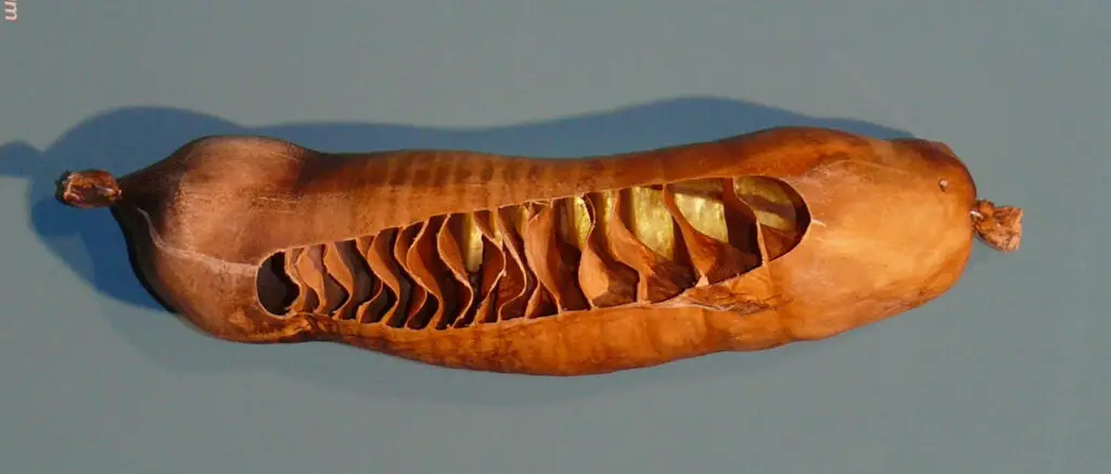
The bursa entiana leads into the straight tubular intestine, bile duct, and pancreatic duct open into the intestine at the starting of the esophagus. In the intestine of Scoliodon, the mucous membrane of the inner wall form a scroll or spiral-like structure, the longitudinal spiral fold is known as the spiral valve or scroll valve.
The spiral fold of the intestine reduces the speed of the flow of food inside the intestine which gives sufficient time for the absorption of nutrients. In Scoliodon the spiral valve is not more than two and a half spirals but in some genera of Dogfish, we can see a large number of spirals in the spiral valve, up to 15 spirals.
Rectum:
The intestine finally meets the short narrow tube of the rectum which opens into the anus, the anus opening outside through the ventral cloaca. In Scoliodon the dorsal wall of the rectum has a finger-like rectal gland but its function is unknown.
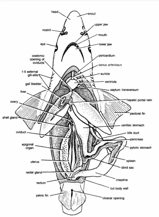
Detailed Study On
Coelom and Viscera in Scoliodon
Placoid Scales In Scoliodon (Exoskeleton)
Skin of Scoliodon (Integument)

Hi Everyone!!! Welcome to Imaluop. Imaluop always try to learn some new and he want to share to other people. Here we will try to learn various topics on Science, specially on Biological Sciences.