The body surface of Scoliodon is very rough, the surface is like sandpaper, on their skin the exoskeletal structure give the roughness, on their skin placoid scales in Scoliodon are arranged in obliquely row and point backwards directions, the placoid scales are dermal denticles, also known as odontoid, these scales are hard and minute tooth like exoskeletal structure.
Structure of Placoid Scales In Scoliodon:
The placoid scales are the tridented sharp calcified tooth-like structures that remain embedded in the skin of Scoliodon and the placoid scales are special features in Scoliodon. A placoid scales have two major components, basal plates and trident spine, the basal plates remain embedded into the dermis layer of skin but the tridented spine remains outside of the skin pointing backward direction.
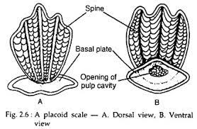
The basal plate is an exoskeletal rhombus-shaped plate that remains embedded into the dermis layer of the integument. The basal plate is made of bone-like loose calcified tissue known as cement and the basal plate is strongly anchored in the dermis by connective tissue fibres. At the centre of the basal plate, the tridented spine arises which project outside through the skin layers.
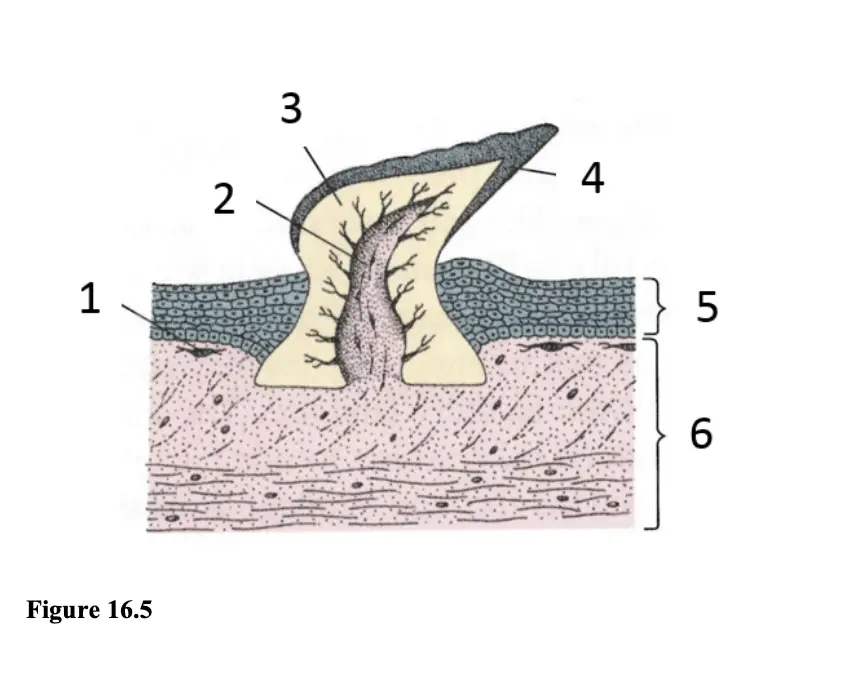
The tridented spine has one median spine and two lateral spines, the tridented spine is hollow and have pulp in the centre which has blood vessels, nerve endings, lymph vessels and some special cells known as odontoblasts. The odontoblasts of the palp produce dentine which forms the wall of the tridented spine. In the dentine of the spine, some network of canals present through which the fine thread-like cytoplasmic process of odontoblasts cells is present.
So internally the tridented spine has a central canal or palp and outside the palp, the wall of the spine is made of dentine, outside the dentine the spine is covered by a hard enamel-like layer known as vitro-dentine.
Development of Placoid Scales In Scoliodon:
The development of the placoid scales starts in the dermis layer of the integument in Scoliodon. In the dermis layer, the development of Placoid Scales start from the development of a dermal papilla, some dermal cells of the dermis layer gather together just below the epidermis and form a dermal papilla.
When the dermal papilla grows more than the upper epidermis layers create pressure on the cells of the dermal papilla. The outermost cells of the dermal papilla turn into odontoblasts, the odontoblasts cells deposit dentine in between the cells of the dermal papilla, at this time the epidermal cells just above the dermal papilla deposit vitro-dentine or enamel on the dermal papilla.
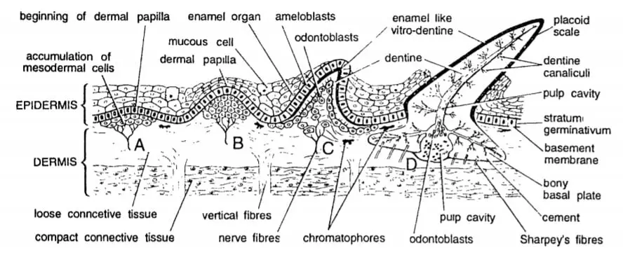
The surrounding cells of the dermal papilla deposit cement which form the basal plate and the dermal papilla develop the palp which has blood vessels, nerve endings and lymph vessels. The palp gives nutrition to the growing dermal papilla, when the dermal papilla develops into a placoid scale, the tridented spine comes outside of the dermis layer but the basal plate remains in the dermis layer.
Homology of Placoid Scales:
If we compare the structure of a placoid scale to the Vertebrate teeth then we can find a close resemblance between them which relates to their homology. The development and structure of Vertebrate teeth is almost similar to the development and structure of a placoid scale so we can consider them as homologous structure.
Even the backwards directed sharp teeth of Scoliodon are actually the modified placoid scales, the teeth in Scoliodon is actually larger and sharper version of the placoid scales present in their skin.
The skin on their body continues into the mouth cavity of Scoliodon and the placoid scales present on their skin specialise in the mouth cavity and become their teeth. So the teeth and placoid scales of Scoliodon are homologous structures and actually, the teeth are only the derivative of the placoid scales.
Previously most of the workers assumed that the placoid scales are both mesodermal and ectodermal because the basal plate and dentine are formed from the mesodermal tissue while the Vitro-dentine is derived from the ectoderm. But now it is assumed that the whole placoid scales are ectodermal in origin.
Detailed Study On
Skin of Scoliodon (Integument)
External Morphology of Scoliodon
Scoliodon: Habits Habitat and Geographical Distribution

Hi Everyone!!! Welcome to Imaluop. Imaluop always try to learn some new and he want to share to other people. Here we will try to learn various topics on Science, specially on Biological Sciences.