The arterial system in Scoliodon has a network of thick-walled muscular arteries, the conus arteriosus of the heart pump the venous blood into the arterial system. The conus arteriosus open into the ventral Aorta, the arterial system start from the ventral aorta. We will discuss all components of the arterial system in Scoliodon with the necessary diagram below divided into some sections for better understanding.
Table of Contents
Arterial System in Scoliodon:
Ventral Aorta:
The heart is present on the ventral side of the body below the pharynx, the conus arteriosus on the anterior side of the body opens into the ventral aorta. The ventral aorta originates from the conus arteriosus and runs mid-ventrally below the pharynx, the ventral aorta reaches up to the hyoid arch and then the ventral aorta bifurcates into two short arteries – the right innominate artery and left innominate artery.
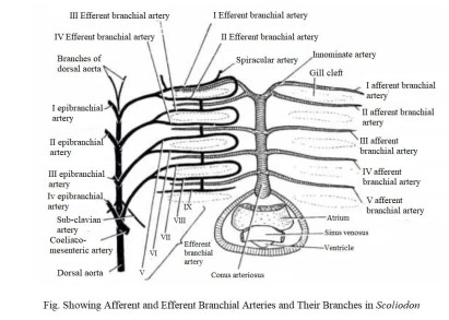
Afferent Branchial Arteries:
Near the hyoid arch each right innominate artery and left innominate artery of them bifurcate again to form the first afferent branchial artery and second afferent branchial artery. At regular intervals third, fourth pairs of afferent arteries originate from the lateral side of the ventral aorta independently. But the fifth pair of branchial arteries do not originate from the ventral aorta independently, the ventral aorta has a median aperture, the fifth pair of afferent branchial arteries arises from the common median aperture of the ventral aorta.
The first pair of afferent branchial arteries run along the posterior side of the hyoid arch and supply blood to the hemibranch of the first pair of the gill. Second, third and fifth pairs of the afferent branchial arteries run through the inter-branchial septa to supply the blood to their corresponding holobranch. When the afferent branchial arteries reach the gill lamellae, the afferent branchial arteries break down into a fine network of capillaries.
Efferent Branchial Arteries:
The afferent branchial arteries form the fine network of capillaries in gill lamellae, all the capillaries join together and form a total of nine pairs of efferent branchial arteries. Each pair of efferent branchial arteries completes a loop around the gill pouches, the anterior portion of the efferent branchial loop is known as pretrematic efferent branchial artery and the posterior part of the efferent branchial loop is known as posttrematic efferent branchial artery.
The first four pairs of efferent branchial arteries make a complete loop around the first four pairs of gill pouches but the ninth efferent branchial arteries have only the anterior portion of the loop. The ninth pair of efferent branchial arteries do not make a complete loop they only present on the anterior side of the last pair of gill pouches.
Epibranchial Arteries:
From the upper region of each efferent branchial artery, loop artery arises known as epibranchial arteries, all the epibranchial arteries join together into a large median dorsal aorta.
Dorsal Aorta:
It is a large major artery that runs along the mid-dorsal line of the body below the vertebral column from the entire trunk to the tail. The dorsal aorta receives oxygenated blood from the epibranchial arteries and supplies oxygenated blood throughout the whole body. During the way of the dorsal aorta, it gives several branches which supply the oxygenated blood to different parts of the body.
- Lateral Aortae: On the anterior end the dorsal aorta, it bifurcate into two arteries, right and left lateral aortae or radix, each of them join with the hyoidean epibranchial arteries of their sides.
- Vertebral Aortae: The dorsal aorta give one pair of branches which supply the oxygenated blood in the vertebral column are known as vertebral aorae.
- Buccal Aortae: The dorsal aorta give one pair of branches which supply oxygenated blood to the roof of the buccal cavity are known as buccal aortae.
- Subclavian Aortae: The dorsal aorta give one pair of branches which responsible for the supply of oxygenated blood in pectoral fins, they are known as subclavian aortae.
- Coeliaco-mesentetic Aortae: Is a single large unpaired artery which gives supply to the stomach, liver, pancreas and intestine.
- Lieno-gastric Aortae: It is a single unpaired artery which gives supply to the stomach, gonads, spleen is known as lieno-gastric aortae.
- Posterior Mesenteric: It also a unpaired branch of the median dorsal aorta which gives supply to the rectal gland.
- Iliac Aortae: One pair of branch arise from the dorsal aorta give supply to the pelvic fins.
- Parietal: In the pathway of dorsal aorta, it gives several small branches which supply blood to the myotomes, gonads, spinal cord, lateral line canal and kidneys.
- Caudal Aortae: The posterior end of the median dorsal aorta continue to the tail region and supply oxygenated blood to the tail region is known as caudal artery.
Hypobranchial Blood Plexus:
On the ventral side of the ventral aorta, one pair of longitudinal small arteries run on the ventral side of the ventral aorta parallelly known as hypobranchial arteries. Two hypobranchial arteries are connected with each other with the help of some transverse blood vessels. Four commissural connect the longitudinal hypobranchial arteries to the lateral network of hypobranchial arteries.
The lateral network of hypobranchial arteries joins the ventral wall of each efferent branchial arteries loop. The two longitudinal hypobranchial arteries finally join together to form a single median coracoid artery which gives two branches – the coronary artery and the pericardial artery. The carotid artery gives supply to the heart and the coracoid artery gives supply to the pericardium, coracoid region of the pectoral girdle.
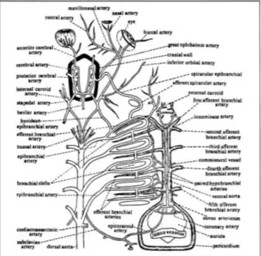
Arteries in Head Region:
Total three branches of hyoidean arteries give supply to the entries head region in Scoliodon, we list all the branches that supply oxygenated blood to the head region.
External Carotid Artery:
The external carotid artery arises ventrolaterally and gives supply to the subcutaneous tissue of the hyoid arch, ventral side of the mandible, coraco-mandibular muscle, superficial hyoid to skin muscle.
Afferent Spiracular Artery:
It continues as spiracular epibranchial which gives a branch to the eyeballs known as the ophthalmic artery, after that it enters into the cranial cavity. Then it joins with the internal carotid artery and is divided into the anterior and posterior cerebral artery which gives supply to the brain.
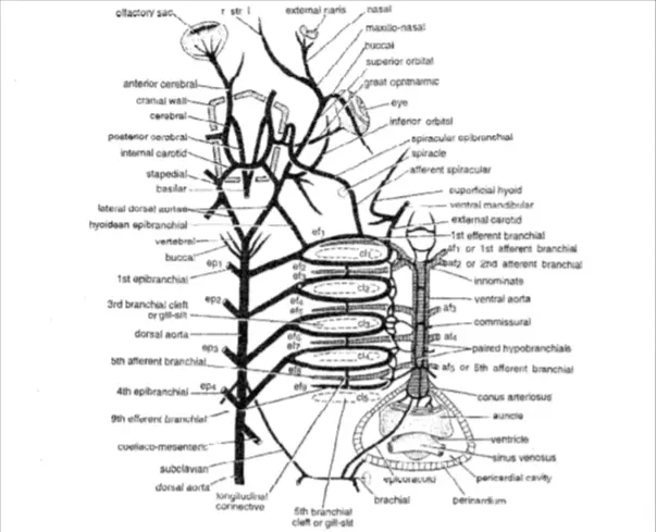
Hyoidean Epibranchial:
It arises from the dorsal wall of hyoidean efferent then on the anterior region they join with the lateral branch of the dorsal aorta and bifurcate into two branches- internal carotid artery and stepadial.
Detailed Study On
Respiratory System of Scoliodon
Digestive Glands and Feeding Mechanism in Scoliodon
Coelom and Viscera in Scoliodon
Placoid Scales In Scoliodon (Exoskeleton)
Skin of Scoliodon (Integument)

Hi Everyone!!! Welcome to Imaluop. Imaluop always try to learn some new and he want to share to other people. Here we will try to learn various topics on Science, specially on Biological Sciences.