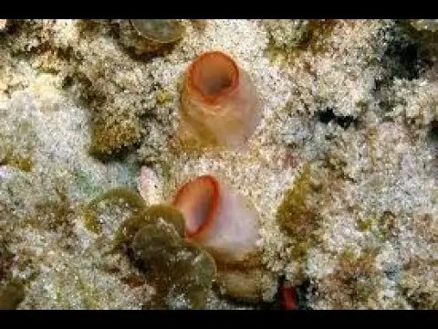
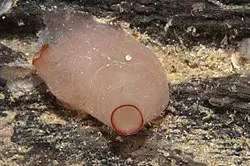
In Herdmania blood vascular system is well developed and it has all of the components of an ideal blood vascular system, a heart with pericardium, blood, and blood vessels.
Table of Contents
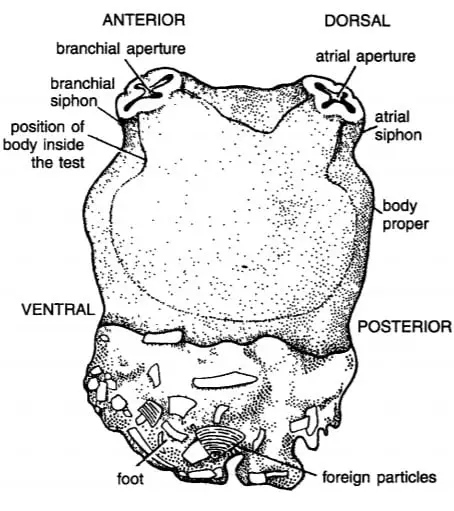
Pericardium:
It is a transparent hollow tubular structure present below the right gonad and is around 7 to 8 cm long and present obliquely. It is a non-contractile structure and the tubular pericardium is closed at both ends, the pericardium is filled with a colorless pericardial fluid.
The pericardial fluid has some corpuscles which show great resemblance with blood corpuscles, the wall of the pericardium is thick made of connective tissue and has blood sinuses, on the interior side, it is lined by squamous epithelium.
Heart:
An infolding of the pericardial wall forms a contractile tubular heart, the tubular heart present inside the lumen of pericardium along the length of the pericardium, both ends of the heart is open. The heart is attached to the wall of the pericardium through a connective tissue flap and the wall of the heart is made of striated muscle. There is no valve in the tubular heart but in the mid-portion of the pericardium, a pear-shaped structure is present which is assumed to control the flow of blood inside the heart.
Blood Vessels:
Herdmania blood vascular system has an advanced vessels system and we listed them which are major blood vessels.
Ventral Aorta:
The largest blood vessel in Herdmania is the ventral vessel or subendostylar vessel which originates from the ventral side of the heart. At the origin, the ventral vessel gives a branch, ventral test vessels which supply blood to the ventral portion of the test, then the ventral vessels are divided into two branches, anterior ventral vessel, and posterior ventral vessel.
Anterior and posterior ventral vessels run in opposite directions, from the anterior and posterior branch of ventral vessels several branches of vessels arises which supply to the wall of the branchial sac, some fine branch supply blood to the mantle and endostyle.
The posterior branch of the ventral vessel gives out branches to the esophagus while the anterior branch of the ventral vessel joins two circular vessels, the peripharyngeal vessel and the sustentacular vessel. The peripharyngeal vessel presents below the peripharyngeal groove while the sustentacular vessel is present at the base of the branchial tentacle.
The circular sustentacular vessel gives 6 to 8 blood vessels to the wall of branchial siphon known as siphonal vessels, the sustentacular vessel gives a branch to each branchial tentacle known as tentacular branch.
Dorsal Aorta:
In the dorsal wall of the branchial sac, above the dorsal lamina dorsal aorta run and it is not connected to the heart directly. But the dorsal aorta is connected to the ventral aorta through some paired transverse vessels and in the anterior portion, it is connected to sustentacular vessels and peripharyngeal vessels.
From the mid-portion of the dorsal aorta a branch supply to the neural complex known as a neural vessel, on the anterior side, it gives a branch to the wall of the branchial siphon.
Branchio-visceral Vessel:
In the posterior region, the dorsal aorta continues to the branchio-visceral vessel which gives two branches, on the right side right branch, or right oesophageal vessel, and on the left side left branch or ventro-intestinal vessel.
The right branch or right oesophageal vessel supplies blood to the right liver lobe and right side of the esophagus while the left branch gives blood supply to the left portion of the esophagus, left liver lobe, stomach, intestine, rectum, and left gonad.
Cardio-visceral Vessel:
On the dorsal side of the heart a vessel arises, a cardio-visceral vessel which gives supply to the right live lobe through the right hepatic vessel, supply blood to the esophagus, and test through the oesophageal-test vessel.
The main dorsal branch gives several branches, a test vessel left the oesophageal vessel, right gonidial vessel, at the base of the atrial siphon it forms a circular sustentacular vessel and 6 to 8 siphonal vessels to the wall of the atrial siphon.
Main middle vessels supply blood to the left gonad through the left gonidial vessel and the main ventral give the dorso-intestinal vessel which supplies blood to the stomach, intestine, and left liver lobe, gastric vessel supply blood to the stomach and the dorsal test vessel supply blood to the dorsal region of the test.
Blood:
Blood is reddish and has colorless and colored blood cells. In blood amoebocyte leucocytes present which are colorless, different types of blood cells with or without nucleus and vacuole. In Herdmania blood special green-colored blood cells are presently known as vanadocytes which are able to extract the vanadium from the seawater and store them inside the vanadocytes.
Reference Habitat and Habits of Herdmania
Detailed Information on
Habitat and Habits of Herdmania
External Morphology of Herdmania

Hi Everyone!!! Welcome to Imaluop. Imaluop always try to learn some new and he want to share to other people. Here we will try to learn various topics on Science, specially on Biological Sciences.