Table of Contents
Primary Excretory Organ:
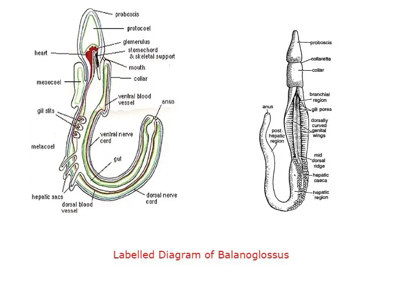
In Hemichordata the proboscis gland or glomerulus is present inside the proboscis coelom, at the anterior portion of the buccal in the Urochordata on the dorsal side of the body above the nerve ganglion the neural gland take part in collecting the nitrogenous excretory material. In Cephaochordata excretion occur through the protonephridia, in each body segment of Cephaochordata protonephridia is present, at the free end of protonephridia a tuft of the solenocytes present, the solenocytes extract the nitrogenous waste materials from the body fluids.
Structure of Excretory Organ:
In Hemichordata the glomerular is a group of tubular structures highly vascularized due to the presence of blood capillaries in the glomerulus. In Urochordata, the neural gland has some peripheral tubules and central tubules. In Cephaochordata the nephridia are L-shaped with a large number of solenocytes on the dorsal side, solenocytes have tentacles like structures in the long stalk-like part and have a rounded nucleated head.
In Hemichordata and in Urochordata there are no solenocytes but in Cephaochordata both anterior and posterior limbs of nephridia have solenocytes.
Physiology of Excretion:
The excretory material present in the blood flow of Hemichordata when reaching in glomerulus glands the nitrogenous excretory material gets collected and excreted into the proboscis coelom. The nitrogenous excretory material gathers in proboscis coelom and finally goes outside of proboscis through the pore present on the anterior end of proboscis, the proboscis pore.
In Urochordata, nephrocytes take part in the extraction of nitrogenous waste materials from body fluids. The nitrogenous excretory material is then discharged into the prebranchial zone.
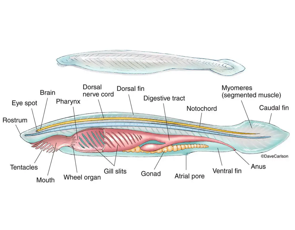
In Cephaochordata protonephridia and the solenocytes remove the nitrogenous excretory material from the body fluids. The solenocytes collect the nitrogenous waste materials from the body fluids then it goes inside the lumen of nephridium, a pore present on nephridia known as nephridiopore discharges the excretory material into the atrial cavity, then the excretory material goes outside of the body through the atriopore.
Secretion of Hormones:
In Hemichordata and Urochordata there are no hormones known associated with the Excretory system. In Cephaochordata the primary excretory organ neural gland also secret some important hormones which control metamorphosis, development in Cephaochordata, neural gland performs similar functions as that of the pituitary gland in the vertebrate.
In Cephaochordata some special structures present associated with the excretory system which is not present in Urochordata and in Hemichordata. In Cephaochordata the Hatschek’s nephridia is a type of nephridia which present at the roof of the buccal hood, in the dorsal region a pair of sac-like structures present known as the brown funnel which takes part in excretion. A group of papillae present in the atrial cavity takes part in excretion known as renal papillae.
Nervous System:
The nervous system in Hemichordata is primitive in nature and it has a great resemblance with the Echinodermata nervous system. In Urochordata, the nervous system in the larval stage undergoes degeneration due to retrogressive metamorphosis and in the adult stage, the nervous system is simple than the larval stage. In Cephaochordata the nervous system is much developed than Hemichordata and Urochordata.
Structure of Nervous System:
In Hemichordata the nervous tissue is present under the epidermis layers, the nerve fibers, and nerve cells in the epidermis from the nerve plexus. The nerve tissue in the epidermis is more concentrated along the mid-dorsal line and mid-ventral line from the dorsal main nerve cord and ventral main nerve cord.
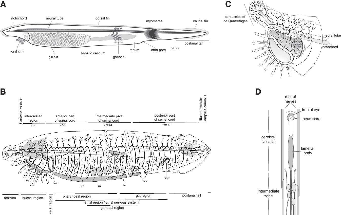
In the Urochordata nerve, the ganglion is present below the neural gland, in Cephaochordata the dorsal hollow nerve cord is filled with cerebrospinal fluid and gives nerve branches in pairs.
Brain:
In Hemichordata no brain-like nervous tissue is present, in Cephaochordata and Urochordata also no brain present but the nerve ganglion in Urochordata and the anterior enlargement on the dorsal nerve cord in Cephaochordata sometimes called as brain by mistake.
In Urochordata and Hemichordata automatic nervous system is absent but in Cephaochordata the automatic nervous system forms the nerve plexus in the intestinal region. In Hemichordata no nerve is present, in Urochordata, some nerves emerged out from the nerve ganglion. In Cephaochordata the nerve cord give several pairs of nerves, first pair of a nerve is known as the cerebral nerve and the other nerves present on the nerve cord is known as the spinal nerve with separate dorsal and ventral roots.
Detailed Study On
Cephalochordata Characteristics Features Affinities Classification Systematic Position
Subphylum Urochordata: Characteristics Classification Examples
Reference
Cephalochordata Characteristics Features Classification Examples and Diagram
Urochordata Classification Morphology Characteristic Features
Characteristics Features of Hemichordata
General Comparison of Hemichordata Urochordata and Cephalochordata
Comparative Study of Digestive System in Hemichordata Cephaochordata and Urochordata

Hi Everyone!!! Welcome to Imaluop. Imaluop always try to learn some new and he want to share to other people. Here we will try to learn various topics on Science, specially on Biological Sciences.